
LASIK and the other refractive surgeries are covered in a number of other websites and I won’t go into much detail here. I’ve included a number of pictures and animations. What is important is that you choose your surgeon carefully. You want a surgeon who is conservative and will tell you if you are not a good candidate. Don’t look for the cheapest, either. If it was my eye, I would get the intralase and I would consider the custom wavefront option if my prescription was -6.00 or higher.
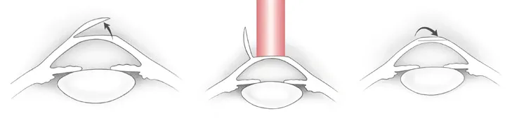
The pictures above show from left to right, the creation of the flap, the laser ablation and the closure of the flap.
Miscellaneous pictures of various retinal pathologies :
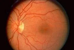
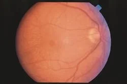
Picture of a normal retina showing the optic disk, the blood vessels and the macula in the center of the photo.
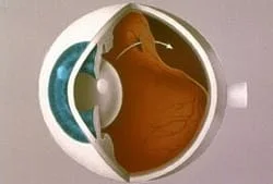
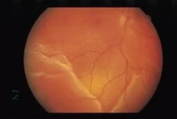
Rhegmatogenous retinal detachment
showing the hole in the inner retinal layer, allowing fluid to separate the inner and outer retinal layers. Unless corrected, this results in total blindness.
Branch Artery Occlusion
showing the emboli blocking the artery just above the optic disk. The bright spot is the emboli.
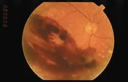
Vitreous Hemorrhage
from diabetes.
Rhegmatogenous (hole present) retinal detachment
.
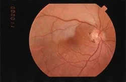
Macular Hole
seen as a round red area in the center of the picture.
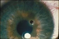
Rusting piece of
metal in the cornea
.
This web site has taken most of its information from the American Academy of Ophthalmology’s Preferred Practice Patterns publications. Each person’s medical condition is unique and all information should be reviewed with their ophthalmologist before deciding on any course of action. We cannot be held responsible for any use, misuse or outcomes from the information contained herein. Thank you.